Hugo J. Kuijf PhD
An experienced researcher, group leader, and educator; with a strong track record in innovative artificial intelligence technology for medical image analysis.
Researcher.
Group leader.
Educator.
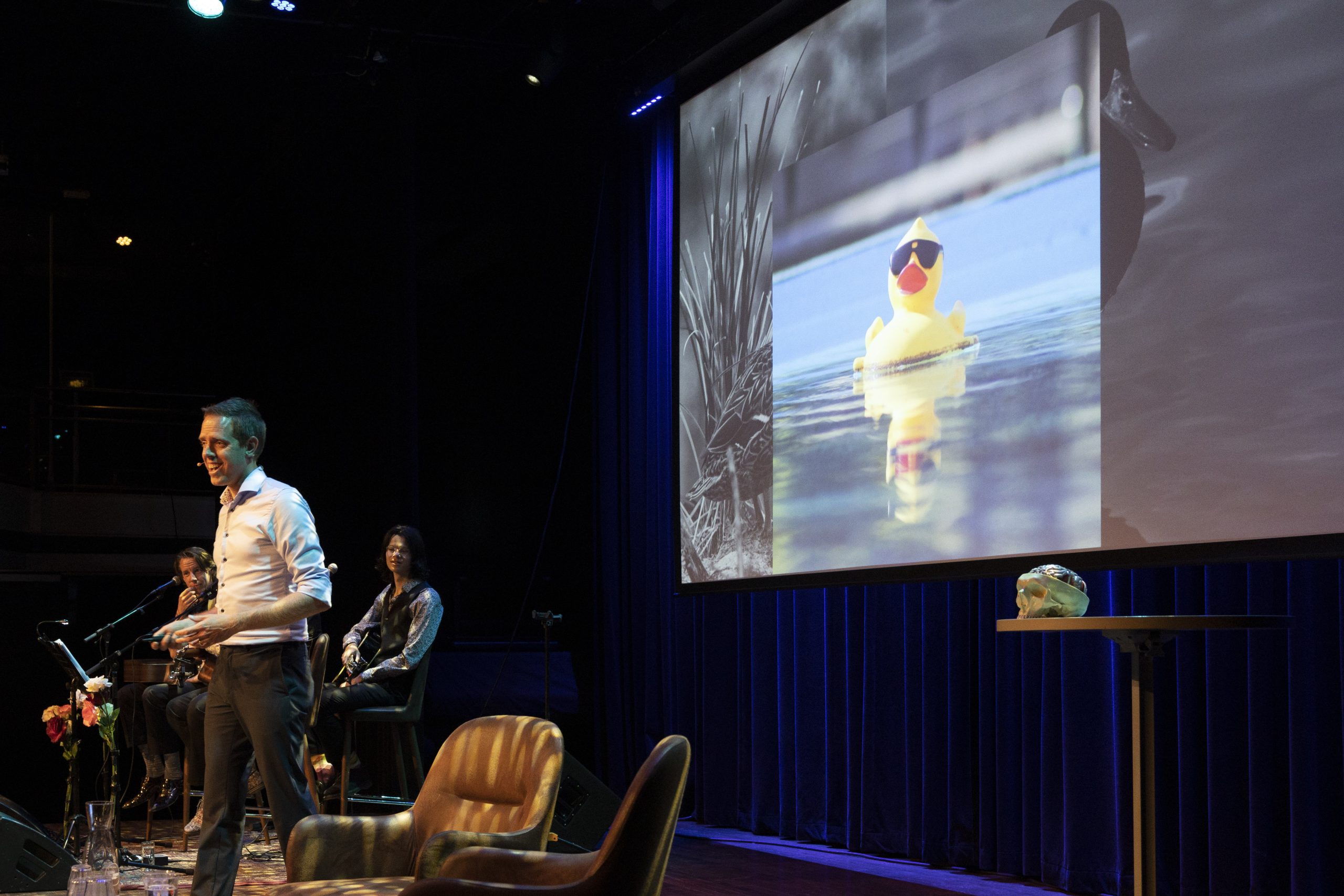
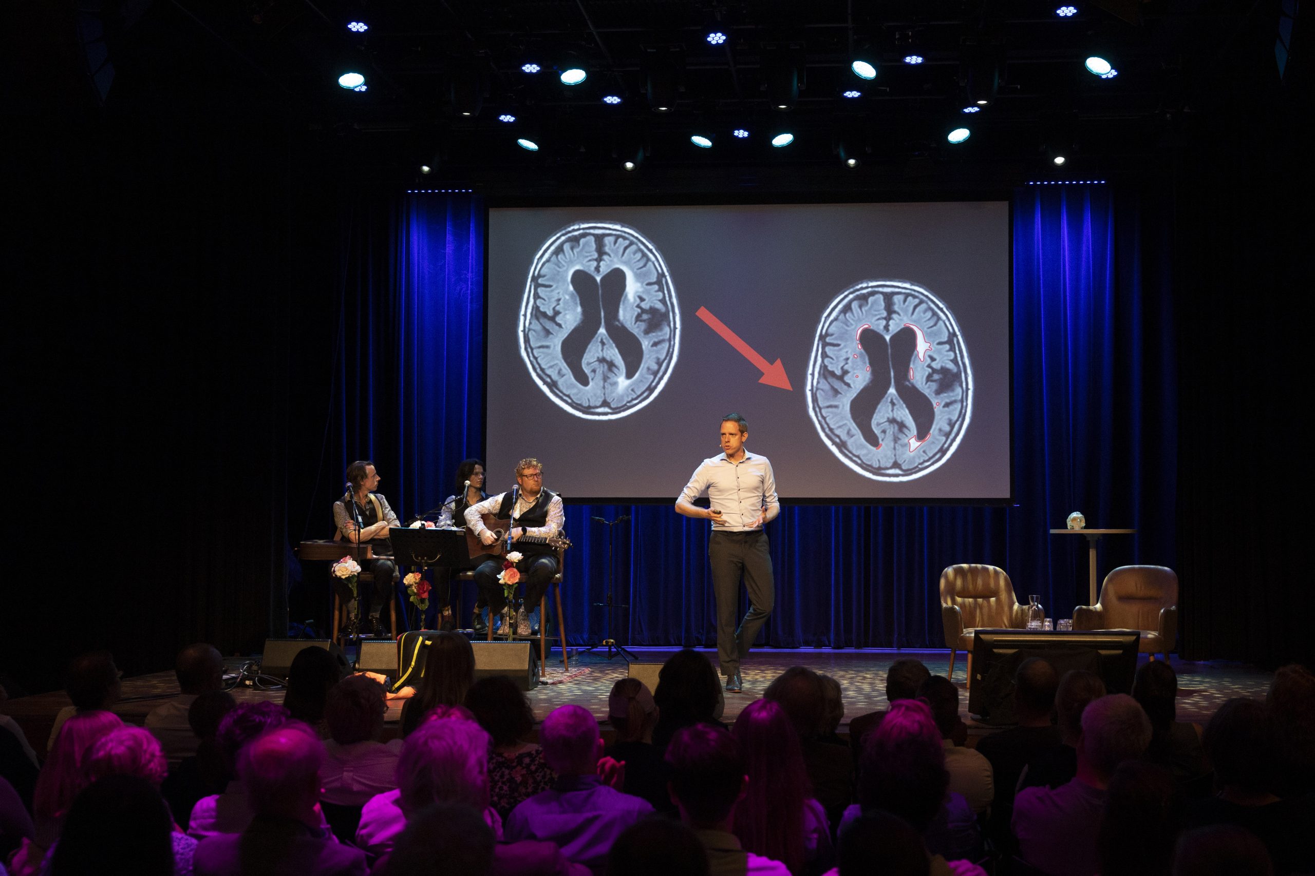
Hugo Kuijf has authored 100+ high-impact scientific papers, published in leading journals. He is regularly invited to speak at international conferences on artificial intelligence in medical image analysis.
He leads a multidisciplinary research group of technical and clinical PhD candidates, working on fundamental innovations in artificial intelligence for medical image analysis and the application in radiology and neurology.
Educating the next generation of scientists in this exciting field is important. Hugo is program coordinator of the Master program Medical Imaging at Utrecht University.

Publications
Publishing both technical and clinical papers allows me to distribute my knowledge and skills to a diverse audience. Supervising early-career scientists in the writing process has been inspiring and insightful.
Speaking
Science communication is a key aspect of my work. I have been invited to speak at well-respected international conferences, provided educational sessions for professional interest groups and students, and explained my work to the general audience.